Definition
Fetal hydronephrosis, (from Greek: ὕδωρ = water, νεφρός = kidney) refers to the dilation of either the fetal renal pelvis alone (pyelectasis or pelviectasis) or of both the fetal renal pelvis and the calices (pelvicaliectasis).
Diagnosis
Seven steps towards establishing the diagnosis:
- Maximum AP RPD (Antero-Posterior Renal Pelvic Diameter) in the transverse plane with spine at 12 (or 6) o’clock.
- Renal parenchyma appearance (normal: iso- or hypoechoic to liver, without cysts).
- Renal parenchyma thickness, which can be measured in the sagittal plane at the level of the mid-kidney as the shortest distance from the renal sinus to the inner aspect of the renal capsule (≥7mm is normal in the third trimester).
- Peripheral calyceal distention in the sagittal plane (abnormal).
- Ureter transverse diameter (≥ 3mm is abnormal).
- Bladder (dilation, wall thickness, or a keyhole sign).
- Amniotic fluid volume (click here to learn more on how to measure the amniotic fluid volume).
After performing the seven steps, categorize your findings according to the UTD system:
| Urinary Tract Dilation (UTD) | 2nd trimester AP RPD | 3rd trimester AP RPD | |
|---|---|---|---|
| A1 | 4mm to <7mm | 7mm to <10mm | central renal pelvis dilation without peripheral calyceal dilation, normal parenchymal thickness and appearance, normal ureters, normal bladder, no unexplained oligohydramnios |
| A2-3 | ≥7mm | ≥10mm | peripheral calyceal dilation, or abnormal parenchymal thickness or appearance, or abnormal ureters, or abnormal bladder, or oligohydramnios from genitourinary cause |
Management
- UTD A1:
- Additional ultrasound at ≥32 weeks.
- Postnatal ultrasound > 48h up to 1 month later. In case of normal findings, repeat examination after 1-6 months.
- UTD A2-3:
- Additional ultrasound initially in 4-6 weeks; certain situations require more expedient follow-up or even consideration of drainage procedures.
- Postnatal ultrasound > 48 up to 1 month later; certain situations require more expedient follow-up.

Unilateral hydronephrosis in a case of megaureter. 
Ureteropelvic junction obstruction. 
Calyceal distention is best imaged in the sagittal plane. 
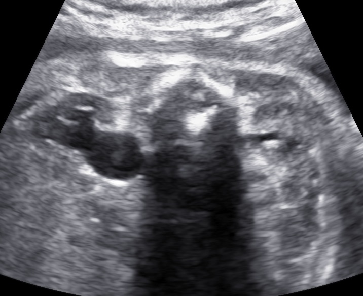
Bilateral hydronephrosis. 
Bilateral duplicated collecting systems; ureterocele and megaureter (postnatal). 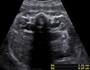
Bilateral hydronephrosis. 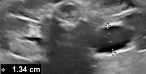
Unilateral hydronephrosis UTD A2-3. 
Normal appearance of the fetal kidneys in the transversal plane at 31 weeks.
Bibliography
- A. Kolitsidakis: fetal hydronephrosis, Visual Encyclopedia of Ultrasound in Obstetrics and Gynecology, www.isuog.org, 27.03.2020.
